Functional geometry of the brain that encode consciousness
Functional geometry of the cortex encodes dimensions of consciousness
Zirui Huang, George A. Mashour & Anthony G. Hudetz
Introduction
Consciousness has been defined conventionally by two separable components: awareness of the environment and the self (i.e., the content of consciousness) and wakefulness (i.e., level of consciousness). For example, patients with unresponsive wakefulness syndrome (UWS)can maintain eye-opening and sleep–wake cycles but are presumably unaware of themselves and their surroundings; thus, their condition is classified as wakefulness without awareness.
The traditional approach to understanding consciousness frequently fails to link specific brain regions to specific functions. The prefrontal cortex, for example, is involved in many functions, such as working memory, decision-making, attention, and task control.
A single brain network can be involved in multiple cognitive processes at the system level. As a result, it is unlikely that isolated brain regions and networks will map onto a single neurofunctional dimension. In the search for neural correlates of consciousness, researchers have typically looked at discrete brain regions rather than continuous brain regions that reflect the brain's intrinsic functional geometry.
The authors hypothesized that consciousness dimensions are encoded in multiple neurofunctional dimensions of the brain, and that a change in consciousness is fundamentally the manifestation of a change along one or more neurofunctional dimensions.
Results
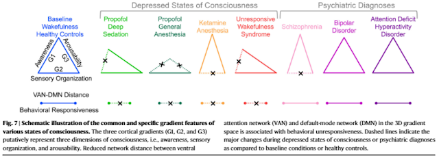
- PDS, PGA, and UWS were associated with a degradation of gradient between unimodal and transmodal areas(Gradient-1)
- KA and SCHZ were associated with a degradation of gradient between visual and somatomotor areas (Gradient-2)
- High-dose propofol (i.e., PGA) further collapsed the functional gradient between visual/default-mode to multiple-demand areas (Gradient-3)
- The authors propose that a given state of consciousness can be represented, independently of cognitive-behavioral description, by a family of cortical gradients derived from the principled characterization of the brain’s functional geometry. Instead of focusing on the presumed role of particular brain regions or networks, this proposed framework may obviate the need to debate if neural correlates of consciousness are, for example, in the front or back of the brain.
- A degradation of the cortical gradient from unimodal to transmodal areas (Gradient-1) may serve as an indicator of disrupted awareness. Furthermore, we found common gradient features in participants with PDS and patients with UWS, i.e., a degradation of Gradient-1 with relatively preserved Gradient-2 and Gradient-3.
The results are consistent with the predictions that both PDS and UWS are associated with disrupted awareness but preserved arousability and sensory organization. - Unlike the heterogeneous clinical conditions often associated with UWS, PDS may serve as a pharmacological model in well-controlled experimental settings. For example, machine-learning models can be trained in a large fMRI database of PDS (with baseline controls) and used to inform clinical diagnosis or prognosis for patients with neuropathological disorders.
- A recent study using a neurobiologically informed whole-brain computational model demonstrated that a change of excitatory-inhibitory balance in favor of inhibition can reproduce brain dynamics that characterize both PDS and UWS. Hence, it is conceivable that an overall increase in neuronal inhibition may be mechanistically relevant to the degradation of cortical gradients, a proposition that warrants future investigation.


Comments
Post a Comment